| Overview:
This lecture introduces some of the special words, phrases and concepts you will need for the course. Just like learning a foreign language - hard at first - but you will find the inhabitants of this new country have some interesting and unusual things to say about human behaviour. Loewi's experiment on vagal nerve stimulation is used to illustrate the beauty associated with the design and execution of a groundbreaking scientific discovery. |
Learning
objectives
|
 Human
brain structure
Human
brain structure

As I mentioned in class, Vesalius was a 14th century anatomist who
spent a good deal of
his time in graveyards and places of execution. He would then dissect
the corpses and make
detailed notes and illustrations. He later commissioned the artist
Titan to do the
illustrations for his book Di Humani Corporis Fabrica .
This book and the
illustrations proved revolutionary in the field of medicine and art.
Vesalius's work is a nice example of how important it is to observe - and accurately report your observations - in any scientific endeavour. Vesalius' approach to anatomy is interesting from a scientist's point of view because he:
| Points to
ponder To what extent does Vesalius' work exemplify the scientific method? How do you think religious authorities in the 14th century would have reacted to the scientific method? |
The next picture shows some parts of the brain that are visible if we slice the human brain in half. As you go through this course you will learn more about the function of these areas, and how they interact with each other to influence our behaviour.
 |
Sections through the brain
I said that the last picture was of a brain that had been
'sliced in half' - not a
very scientific way of talking about things! There are several ways you
could slice a
brain in half and not end up with the view in the picture. To get
around this problem, and
make it clear how the brain was sliced, we need to introduce the
terminology used by
anatomists when they section the brain. This is an example of an operational
definition
- putting an operation into a form of words that can be understood by
everyone.
 |
You create a coronal section when you slice bread |  |
| You create a sagittal
section when you cut an apple in half |
||
| |
You create a horizontal section when you cut a bread roll to make a beefburger |
Here is a midsagittal
section ( = a sagittal section through the midline) of rat brain from
the UCLA Laboratory
of NeuroImaging 
The most important point about this picture is that the structures we
explored in the
human brain are also found in the brains of rats as well as other
animals.
This is the reason why psychologists are so interested in rats. It is obviously impossible to interfere with the human brain to understand how it works. Although we can gain valuable insight into our brain by observing the consequences of illness and accidental damage, these tragic events often involve more than one area of the brain.
Animals are used because it is possible to control the extent of a lesion or area of stimulation under laboratory conditions.
Some of you may be concerned with the ethical issues surrounding the use of animals in this way. Carlson (2001) puts forward the case for using animals to alleviate human suffering.
More
details on a rat brain atlas available
over the Internet are given in the Supplementary Information section at
the end of these
notes
| Point to ponder
Why do you think people bother to make detailed maps of rat brains? |
Directional terms
Now that we have seen how to cut the brain to reveal its structures, we
need to describe
the way in which anatomists use general terms to describe the relative
position of
structures to each other. Anatomists use the following words to
describe direction
in the brain and other parts of the body:
| Direction | Description | |
| Ventral | Toward
the front (belly) of the body or towards the bottom of the head |
|
| Dorsal | Toward
the back of the body, or towards the top of the head |
|
| Rostral | Toward the nose | |
| Caudal | Toward the feet (humans) or tail | |
| Lateral | Away from the midline | |
| Medial | Toward the midline | |
| Bilateral | On both sides of the body or head | |
| Ipsilateral | On the same side of the body or head | |
| Contralateral | On the opposite side of the body or head |

|
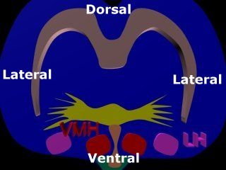 |
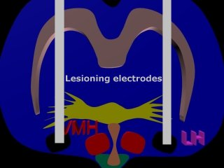
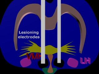
It is important to
clearly distinguish between parts of the brain that are very close
together - they can often have very different functions!
The diagram shows the location of the lateral hypothalamus (LH) and ventromedial hypothalamus (VMH) in a coronal section of the rat brain. Notice how the names of these brain areas reflect their relative positions in the brain.
This diagram also illustrates bilateral symmetry in the brain. Each side of the brain is a mirror image of the other side |
 |
 |
Neuron- a cell specialized for information transmission
 |
If you were to
enormously magnify the pictures of brain sections we have examined in
this lecture you would eventually be able to see neurones.
Neurones are brain cells that specialize in communication. The brain contains circuits of interconnected neurones that pass information between themselves. The picture of a raised hand is an easy way to remember the relationship between:
In neurones, information passes from dendrites through the cell body and down the axon. This is easy to remember because when you pick up an object, the sensation travels from your fingers through your hand, and down your arm. Transmission of information through the neurone is an electrical process. The passage of a nerve impulse starts at a dendrite, it then travels through the cell body, down the axon to an axon terminal. Axon terminals lie close to the dendrites of neighbouring neurones. When the nerve impulse reaches an axon terminal it causes the release of a chemical ( called a neurotransmitter ) that travels across the gap (the synapse) between a terminal and the dendrite of the neighbouring neurone. Neurotransmitters stick to receptors in the neighbouring dendrite and trigger a nerve impulse that travels down the dendrite, across the cell body, down the axon etc. Our behaviour is the consequence of millions of cells talking to each other via these chemical and electrical processes.
|
 |
The
synaptic junction between neurones

1. Bridging the
information gap between neurones
Neurotransmitters are responsible for transmitting information across the synaptic gap between neurones.
Neurotransmitters are stored in synaptic vesicles. When action potentials are conducted down an axon:
Neurotransmitters in the synaptic cleft :
 |
2. 'Mopping up'
after information transmission
This
image illustrates the main events thought to be involved after
transmitters have been released into the synaptic cleft.
|
| Point to ponder
Why do you think some neurotransmitter is broken down by enzymes and some undergoes reuptake? |
Loewi's experiment on
vagal nerve stimulation
We will now examine a groundbreaking discovery by Loewi in 1921, that
shows that neurotransmission
involves the release of a chemical which transmits
information between neurones
|
Working in 1921, Loewi knew that electrical stimulation of a frog's
heart stopped heart
contractions. He suspected that a chemical was released when the nerve
was stimulated,
because this effect persisted for a short time after the stimulation
ceased.
At 3a.m. one night he had a flash of insight into how he could test this idea and he went to the lab and carried out the experiment that made him famous, and demonstrated for the first time that neurotransmission involved the release of a chemical substance.
In a
later experiment (illustrated here) the
sympathetic accelerator nerve of the donor heart was stimulated,
producing increased heart
rate. The donor heart released epinephrine which - a few
seconds later - increased
heart rate in the recipient heart. These experimental finding lies at
the foundation of
all modern medicines used to treat mental illness.
The cardiac branch of the vagus nerve attached to a frog's donor heart
was electrically
stimulated, causing arrest of the heartbeat. Ringers solution perfusing
the donor heart
was transferred to the recipient heart. After 15 seconds, the beat in
the recipient heart
was also arrested. Loewi's experiment is a nice example of how
important hunches are in
experimental design.
| Point to ponder
What does Loewi's experiment tell us about communication between cells in the brain ? |
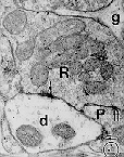
Electron microscope pictures of synapses
 |
 |
Here
are some pictures of synapses taken with
an electron microscope and Eric Chudler's description of them:
"Developed in the 1950's, the electron microscope can magnify objects
thousands of
times. Electrons are passed through an object and then recorded on
film.
Using the electron microscope, Dr. Pati Irish in the Department of
Neurological Surgery at
the University of Washington has taken these pictures of synapses. The
"d"
represents a dendrite and the "R" represents an axon terminal. If you
look
closely, you can even see some round synaptic vesicles that contain
neurotransmitters. The
fuzzy black areas represent the actual synapse between terminal and
dendrite. The larger
oval objects (there are two in the dendrite of image 1 and one in the
dendrite of image 2
are 'mitochondria'. "
There is a link to Eric Chudler's description of synaptic events , and pictures of synapses using the electron microscope in the Supplementary Information section at the end of this page - well worth a visit.
I have written a Java applet that allows you to magnify these images so that you can examine the detail. The horizontal and vertical scroll bars enable you to move around the image.
| Supplementary
Information Neurotransmission
Brain anatomy
|