Author Paul Kenyon
Nerve Cells and Nerve Impulses
If you cannot see the VRML model on the left,use the National Institute of Standards and Technology website
to automatically detect the correct VRML plugin for your browser.
|
VRML model |
Author Paul Kenyon Nerve Cells and Nerve ImpulsesIf you cannot see the VRML model on the left,use the National Institute of Standards and Technology website to automatically detect the correct VRML plugin for your browser. |
"Behaviour is the consequence of the activity of cells in the brain called neurons." Now there is a bold statement for you! One fundamental feature of behaviour is its variability. Sometimes I eat because I am hungry, sometimes because I am bored and often because the food is just irresistible. The last sentence stated that 'I' made the decision to eat. What is this 'I'? One explanation is that 'I' is a collection of nerve cells with the ability to perceive, remember and act. This type of explanation is sometimes called 'reductionist' because it reduces behaviour to interactions between (apparently) simple processes. You need to be aware that reductionists come in for a fair amount of criticism because reductionism appears to rob us of our 'free will'. These are important philosophical questions. You won't get a complete answer in this set of lectures but you will hopefully be in a better position to appreciate the strengths and weaknesses of the various arguments by the end of the course. In this section we begin to explore some of the ways in which brain cells communicate with each other.
 | The human body consists of billions of cells. Cells in different parts
of the body are specialized - blood cells carry
oxygen, stomach cells absorb food, kidney cells produce urine, bone
cells form the skeleton, muscle cells move our joints etc. Neurons are brain cells that specialize in communication. The brain contains circuits of interconnected neurons that pass information between themselves -they are the main decision makers in the body. |
|
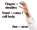
Here is a picture showing a simple way to remember
the various parts of a neuron and the direction of information
transmission.
|
The passage of a nerve impulse through a neuron starts at a dendrite, it then travels through the cell body, down the axon to an axon terminal. Axon terminals lie close to the dendrites of neighboring neurons. When the nerve impulse reaches an axon terminal it causes the release of a chemical ( called a neurotransmitter ) that travels across the gap (the synapse) between a terminal and the dendrite of the neighboring neuron. Neurotransmitters stick to receptors in the neighboring dendrite and trigger a nerve impulse that travels down the dendrite, across the cell body, down the axon etc. Our behaviour is the consequence of millions of cells talking to each other via these chemical and electrical processes.
Electrical events in neurotransmission
The outer 'wall' or cell membrane of neurons plays
a vital role in their ability to transmit electrical impulses. It is semi-permeable
which means that electrically charged chemicals called ions
can pass in and out of the neuron. Normally the concentration of ions
is different inside and outside the neuron. This causes an electrical
difference between the inside and outside of the neuron. We can measure
the electrical state of a neuron by placing a tiny electrode inside the
neuron (see Figure 5.1 in Rosenzweig et al ). This electrical property
is called the resting or membrane
potential.
The inside of the neuron is electrically negative (about -70
millivolts) with respect to the outside. When
neurotransmitters attach to receptor sites on a neuron's dendrite they
can increase (hyperpolarization) or decrease (depolarization)
membrane potential. During
depolarization membrane potential decreases from -70 millivolts. If
depolarization reaches -50 millivolts an action potential is
triggered. During the action potential the membrane
potential reaches +40 millivolts. Triggering an
action potential is like squeezing the trigger on a gun. If enough
pressure is applied the gun will fire. Both a gun and a neuron have a threshold
at which they will fire. Both events are 'all-or-none'.
If too little pressure/depolarization is applied, neither the gun or
the neuron will fire. But once the threshold is reached, increasing
pressure/depolarization has no further effect. In fact, the membrane
potential the action potential dies away and the membrane returns to
its resting state of -70 millivolts. Figure 5.2 in Rosenzweig et al
illustrates the effects of depolarizing stimuli on the electrical
properties of a neuron. This discussion has focussed on explaining how
the combined excitatory postsynaptic potentials (EPSP) produced many
excitatory synapses
At this point we have seen how information is passed from one neuron to
another neighboring neuron. Next we need to examine how this
information is passed down the neuron so that the information can be
propagated through a neural circuit. The action potential passes down
the dendrite and across the cell body as a wave of
depolarization. This is a relatively slow process but adequate because
the distances involved are very small. But the picture changes when we
come to transmission down the axon.
Recall that axons are relatively long structures. For example, axons
are several meters long in the giraffe, and individual axons carry
messages from your spinal cord to muscles in your feet. The conduction
speed of impulses down axons is a function of their size:
the thicker the axon the faster conduction occurs. In addition there
are two types of axon: unmyelinated and myelinated.
Myelin is a fatty sheath that covers the outside of the axon. There are
gaps in this sheath called nodes of Ranvier. Action
potentials jump from node to node in myelinated
axons. This is called saltatory
conduction - from the Latin word saltus meaning
leap or jump; the word somersault comes from the same root. Saltary
conduction is very fast; Rosenzweig et al give the following figures:
| Unmyelinated | Myelinated | |
| Axon diameter | ||
| 1 mm | 2 meters / second | |
| 2 mm | 5 meters / second | |
| 20 mm | 120 meters / second |
 |
If the human brain did not contain myelinated axons it would have to be 10 times bigger than it is to maintain the same velocity of nerve impulses. It makes you wonder about ET! |
Behaviour is the result of excitatory and inhibitory neurons
This discussion has focussed on explaining how one neuron triggers an electrical response in an adjacent neuron. But a moment's reflection reveals that a brain consisting of a network based on this type of information transmission would have limited control over behaviour. Imagine a network with just three neurons :A, B and C;
Clearly neuron B adds very little value to the network. it could be eliminated with no loss in functionality.
There are four things that introduce tremendous flexibility into neural networks:
Recall that when neurotransmitters attach to receptor sites on a neuron's dendrite they can increase (hyperpolarization) or decrease (depolarization) membrane potential. [A hyperpolarized membrane potential is one that is greater than the resting level (-70 millivolts) e.g. -80 millivolts.] This means that that a neuron can have an inhibitory (mediated via hyperpolarization ) as well as excitatory (mediated via depolarization) effect on a neighboring neuron.
We have seen that during depolarization, membrane potential decreases from -70 millivolts. If depolarization reaches -50 millivolts an action potential is triggered. But if an action potential is not triggered (say it only reaches -60 millivolts), membrane potential returns over a short period of time to the resting potential of -70 millivolts. Thus there is a short 'window of opportunity' during which a second weak stimulus can build upon the already reduced membrane potential to trigger the full blown action potential. This is called temporal summation.
These subthreshold events can be caused by inhibitory (inhibitory postsynaptic potentials, IPSP) as well as excitatory (excitatory postsynaptic potentials, EPSP) inputs to a neuron. This table sets out the relationships between the various terms:
| Excitatory input | Inhibitory input | |
| Effect on resting membrane potential (-70 millivolts) |
depolarization = reduced membrane potential | hyperpolarization = increased membrane potential |
| Weak effect | excitatory postsynaptic potentials, EPSP e.g. -60 millivolts | inhibitory postsynaptic potentials, IPSP e.g. -80 millivolts |
| Strong effect | excitatory postsynaptic potentials, EPSP of -50 millivolts triggers action potential | inhibitory postsynaptic potentials, IPSP e.g. -90
millivolts. No action potential |
Spatial summation refers to the finding that several inputs (excitatory or inhibitory )originating from separate locations can exert a cumulative effect on a neuron.
Use the viewpoints (by clicking click the > key underneath the model) for the VRML model on this page to familiarize yourself with the following parts of a neuron
The model contains three neurons which we can name for the sake of convenience
which make synaptic connections with the dendrites of a single neuron which we can call Action!
When Action! fires, the dog retrieves the ball.
As you explore the model try to construct a mental picture of how the Fetch!, Throw Ball and Distracting Dog neurons interact to influence your dog's retrieving behaviour.
Triggering the Throw Ball neuron does not elicit an action potential in the Action! neuron.
Triggering the Fetch! neuron elicits a short lasting excitatory postsynaptic potential rather than a full blown action potential in the Action! neuron
There is a short 'window of opportunity' during which the membrane potential in the Action! neuron is depolarized. During this period the Throw Ball neuron can build upon the reduced membrane potential to trigger an action potential in the Action! neuron. Follow these steps to observe this effect:
Your dog fetches the ball and returns it to your hand.
Follow these steps to see how shortlasting this effect is:
This time - because you waited for the effect of the excitatory
postsynaptic potential to disappear - your dog does not chase and
retrieve the ball.
Perform these steps several times until the effect of imposing
a delay between triggering the Fetch! and Throw Ball neurons
is clear to you.
Inhibitory neurons hyperpolarize
the membrane of neighboring neurons and make them resistant to the
effects of excitatory input.
The Distracting Dog neuron is an inhibitory neuron which hyperpolarizes
the Action! neuron.
Explore the effect of IPSP on EPSP
Observe that the Action! neuron does not fire and your dog does not fetch the ball. You need to overcome the hyperpolarization in the Action! neuron by triggering the Fetch! neuron a second time. Thus if you now
the Action! neuron fires, and your dog retrieves the ball.